
3D x-rays are used often for dental implant treatment. They help me increase the safety and precision of your dental implant surgery. 3D x-rays are often called CT scans, 3D scans, or cone beam scans. This can be done at an x-ray lab or at my office in Burbank.
Cone beam 3D scans are extremely safe and have very little radiation. In my office, I have a very technologically advanced machine that can take a limited view scan. This is called a “limited field of view.” The big advantage to you is that the radiation can be focused to only one tooth! In the past, if you wanted to see one tooth area, the whole jaw or head had to be x-rayed.
With a cone-beam rather than a medical “spiral CT,” the beam is extremely focused and 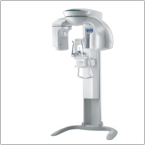
My cone beam machine looks like this and takes most scans in about 30 seconds:
The safety and precision of the 3D scan allows me to do virtual surgery on my laptop before I ever do it in your mouth! This reduces the number of surprises that can be encountered during surgery. It allows me to see your nerves, bone arteries, sinuses, bone depressions, and every detail down to less than a 10th of a millimeter. The detail of virtual reality is amazing.
Having this technology in the office prevents you from having to drive all over town from the x-ray lab, to the dental implant surgeon, to the regular dentist that will make the tooth. In my office, all of these are in my control, under one roof. This allows me to offer dental implants and teeth safely, at a fair price, and most of all, to do it right.
Another big advantage is often a bone graft can be avoided if a 3D scan is done. Even if the bone is narrow, the trajectory can be changed and/or an angled custom abutment can be used.
A 3D x-ray is not required for all dental implants and bone graft procedures. I asses each patient carefully before recommending a 3D cone beam x-ray. Some patients require two or more 3D x-rays– one before major bone grafting and one after.
Ramsey A. Amin, D.D.S.
Diplomate of the American Board of Oral Implantology/Implant Dentistry
Fellow-American Academy of Implant Dentistry
Burbank, CA
Great blog. VERY informative! I wish I lived near you!
Bill
Cone beam technology is an important breakthrough for dental implant therapy.This technology has more benefits for patients. Good information shared!I take pleasure in learning about this topic.